Macular degeneration
Age-related macular degeneration is the most common cause of irreversible loss of vision in the western world. This disease affects the central area of the eye’s fundus, which is the most important area. What is the result of this ? A gradual reduction of central vision with no other symptoms.
What is macular degeneration?
 Several studies have calculated that approximately 6% to 10% of people between the age of 65 and 74 and about 19% to 30% of people over the age of 75 have this disease. As we can clearly see it is directly related to advanced age and for this reason it is called macular degeneration of the elderly.
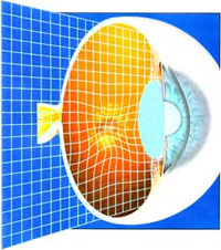
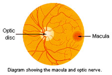
The macula corresponds to the central part of the retina which is the internal part of the eye. This area is the most important area of the retina on which rays fall and the image is formed. It is understood that the macula is the most important part of the retina for the maintenance of clear eyesight..
What causes macular degeneration?
Age related macular degeneration is a multi-factorial disease ie. it has been proved that several factors play an important role in its appearance. These risk factors may be age, heredity, light-colored irises, smoking, cardiovascular conditions, as well as sunlight. Naturally, the most important factor is ageing.
Symptoms. Clinical picture
Age-related macular degeneration is a disease whose symptom is the gradual reduction of central vision. There are no other symptoms such as pain. Vision is never totally lost since peripheral vision remains. So our central vision is no longer clear when we look at an object, but we can still move around in a room (orientation) because our peripheral visual field remains normal. However, in advanced cases we have a real disability since our vision is extremely poor.
There are two types of macular degeneration – dry (atrophic) which refers to 90% of cases and wet (exudative) referring to 10% of patients. The latter is however responsible for 90% of considerable vision loss in patients with degeneration. In these cases, new abnormal vessels can appear under the retina, forming a coat under the macula. As a result vision is decreasing.
What can we do to prevent it?
- Wear sunglasses with UV filter to protect your eyes.
- Take food supplements, multi-vitamin compounds and zinc compounds. Although it is hard to prove these medicines’preventive action, several studies have shown that they can help delay the disease’s onset. Your ophthalmologist will advise you on the correct dosage, after consulting with a pathologist for possible contraindications.
- Have preventive ophthalmological check-ups after the age of 40 and visit your ophthalmologist as soon as you notice changes in your vision, particularly shadows in your central eyesight.
- Reduce, or better yet, quit smoking. Besides it is known how harmful it is, not only to the eyes, but also to the rest of the body.
- Control your pressure and cholesterol. Should there be an indication, visit a cardiologist.
Special diagnostic examinations
 What we must know is that we should undergo periodical screening tests, and if necessary, have a special examination where the fundus of the eye is photographed, following the administration of a special dye, fluoroscein. This test is called
fluoroscein angiography. Through this test we are able to determine whether there are lesions in the retina and the macula and whether abnormal vessels are formed under them. This diagnostic method is undertaken through the administration of fluoroscein in the blood. This makes photographing abnormal vessels and their exact dimensions possible.
What we must know is that we should undergo periodical screening tests, and if necessary, have a special examination where the fundus of the eye is photographed, following the administration of a special dye, fluoroscein. This test is called
fluoroscein angiography. Through this test we are able to determine whether there are lesions in the retina and the macula and whether abnormal vessels are formed under them. This diagnostic method is undertaken through the administration of fluoroscein in the blood. This makes photographing abnormal vessels and their exact dimensions possible.
Also, another test is macular tomography (OCT-optical coherence tomography) during which the retina is examined, layer per layer, and lesions on the macula can be seen without having to inject dye and no pupil dilation is needed. OCT is used to monitor the course of treatment and the evolution of degeneration.
Treatment
In case of dry macular degeneration, there is no treatment. Disease progresses slowly but we cannot prescribe pharmaceutical or surgical treatment which can solve the problem. In this case, the patient can take multi-vitamin food supplements which include lutein and zeaxanthine.
In case of wet macular degeneration and if there is a neovascular membrane present, then treatment of choice, from 2005 until today, is the administration of antiVGEF drugs in the form of intraocular injections. These medicines (AVASTIN, LUCENTIS) act in order to block the neovascular membrane and diminish or completely wipe out the swelling (fluid) in the area of the macula. As a result, degeneration is not only stabilized and restrained, but many times, the patient’s visual acuity is improved. These injectable medicines are administered every 4-8 weeks until the disease is fully stopped.
In special cases, macular degeneration can be surgically treated. Macular translocation is an operation which is recommended for patients whose vision is completely lost in both eyes due to macular degeneration. With macular translocation we are able to move the macula to a new healthy spot in order to regain functional vision in one eye.
Low vision aids
In the case of advanced degeneration, low vision aids can also be used. These are special glasses or special magnifying lenses or closed television circuits and computers which help low vision by magnifying objects or the visual field. These are aids which only the ophthalmologist can recommend and the patient is trained to use them first. However, it must be pointed out that these represent an important solution to a difficult problem, particularly when all our other therapeutic weapons have failed.