The Keratoconus
Keratoconus is a relatively common eye disease, extensive, non-inflammatory central dystrophy of the cornea. The cornea takes on a conical shape, causing an abnormally progressive astigmatism resulting in blurred vision. Its aetiology can be due to a number of factors, including heredity, biochemical abnormalities, and self-induced mechanical trauma to the cornea. The treatment remains the use of hard contact lenses. A minority of patients need corneal surgery, ie a corneal transplantation to regain vision when other treatments do not have the desired effect.
 Introduction-Pathogenesis
Keratoconus is a dystrophy in which the cornea takes on a conical shape. The result is an abnormal astigmatism with consequent impaired and blurred vision. The disease affects both eyes and occurs between the ages of 10 and 30 years.
Keratoconus is considered a multifactorial disease. Many studies converge on the view that keratoconus is due to metabolic abnormalities of the cornea. The association of the disease with other metabolic diseases supports this view.
The heredity factor has not yet been determined. However, this theory is supported by the fact that twin siblings display a high percentage of keratoconus. Recent studies show that patients with a positive family history develop keratoconus in 6% -8% of cases. In any case, a child of parents with keratoconus has a 1/100 chance of developing the same disease.
One of the common causes of keratoconus is the mechanical trauma caused by the intense "rubbing" of the eyes with our hands. An association of the disease with allergic tendencies and eczema in the eye area has been found. This mechanical theory is also confirmed by the fact that many patients with keratoconus have soft eyelids causeing them to turn back easily. Down syndrome is also another risk factor for developing the disease.
Diagnosis
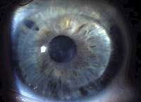
In the early stages, the patient complains of a reduced visual acuity. Physical examination does not always provide evidence for a definitive diagnosis. However, keratometry can reveal altered parameters. The patient presents with an abnormal progressive astigmatism that he did not have before. In more advanced stages, the diagnosis is easier and with the help of the slit lamp the cornea shows the familiar conical shape as well as thinning and blurring of its central area.
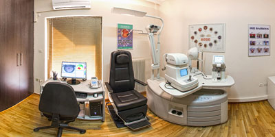
The definite diagnosis, however, is made with the help of an electronic machine and with an examination called “corneal topography” (corneal mapping). With this examination we obtain a two-dimensional colour imaging of the topography of the cornea on the basis of which we make a diagnosis even in subclinical forms, ie those that have not shown symptoms.
Treatment
In early keratoconus, correcting with glasses for astigmatism is the easiest way to treat it. However, the main treatment for keratoconus is the application of semi-rigid contact lenses, which alter its abnormal conical curvature. Soft lenses are not indicated as they do not dramatically change the curvature of the cornea.
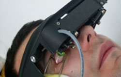 Collagen cross-linking (CxL) is considered to be the newest method of surgical treatment of keratoconus. This method uses ultraviolet radiation (30 minutes of exposure of the eye to the lamp) and simultaneous instillation of a drug, riboflavin, in drops. In this way a "self-aging" is caused in the cornea and this has the effect of stabilizing the cone in the phase in which it is. Suitable for onset keratoconus, in young people (under 30 years) and in corneas without advanced thinning (over 400 μm thick)
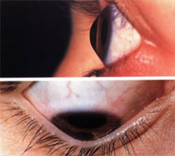
Surgery is needed in about 10% of patients. The operation that is performed is penetrating keratoplasty or corneal transplant in which the patient’s cornea is replaced by another normal one. The operation is performed under local or general anesthesia as an outpatient. The results are very good. A visual acuity of 5/10 postoperatively reaches 90%. 60% of patients need contact lenses postoperatively due to the astigmatism caused by suturing. Sutures are usually removed after 1 year.